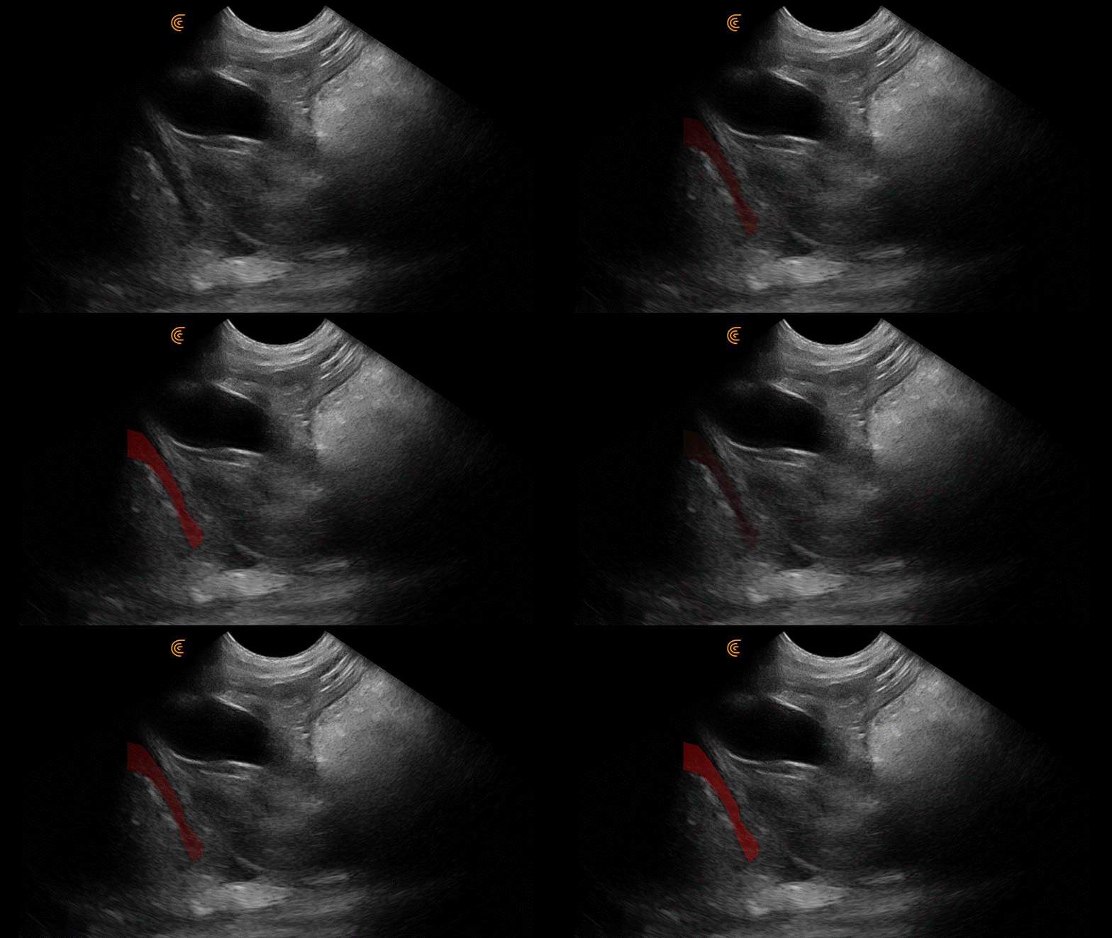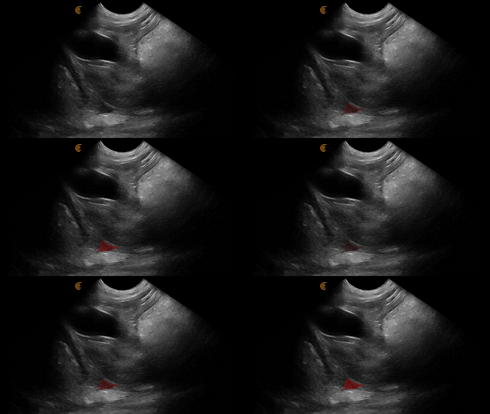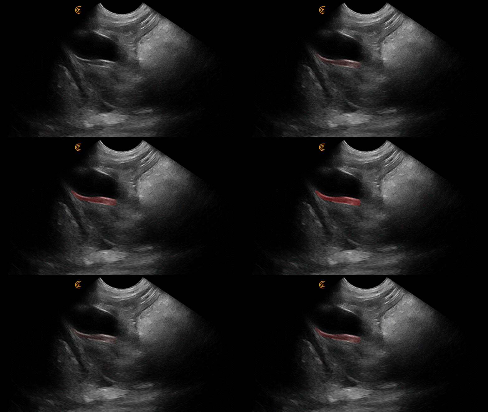
The subxiphoid (DH) view can provide a ton of information. This screen demonstrates the presence of pericardial effusion, pleural effusion, and the gallbladder halo sign.
Tip #1: If the heart kisses the diaphragm, significant pericardial effusion is unlikely. Here, the heart is separated from the diaphragm due to effusion.

Tip #2: Pleural effusion vs pericardial effusion? Pleural effusion will track along the diaphragm creating distinct triangles.

Tip #3: The gallbladder halo sign is caused by more than anaphylaxis. Any process leading to hepatic venous congestion can lead to gallbladder wall edema and include right-sided heart failure and pericardial effusion. Other reported causes include pancreatitis, low albumin, and immune-mediated hemolytic anemia.

Leave a Reply